Australia has one of the highest rates of skin cancer in the world. The combination of intense ultraviolet (UV) radiation, outdoor lifestyles and fair skin means that approximately two out of every three Australians will develop some form of skin cancer during their lifetime. Fortunately, the vast majority of skin cancers can be successfully treated when detected early.
Skin cancer develops when UV radiation damages the DNA of skin cells. Over many years this damage accumulates, causing abnormal cells to grow uncontrollably.
At Skin Cancer Laser Centre, our philosophy is simple: early detection, effective treatment, skin cancer prevention, and achieving the best possible cosmetic outcome.
The Three Major Skin Cancers
Basal Cell Carcinoma (BCC)
The most common skin cancer. Usually slow-growing and rarely spreads, but can cause significant local destruction if left untreated.
Squamous Cell Carcinoma (SCC)
More aggressive than BCC — has greater potential to invade deeply and to spread to lymph nodes, especially when high-risk features are present.
Melanoma
The most serious form of skin cancer because it has the greatest potential to spread. Early-stage melanoma is highly treatable — which is why regular checks matter.
On this page
Melanoma
What is Melanoma?
Melanoma is a cancer that develops from melanocytes, the pigment-producing cells that give our skin its colour. Although melanoma accounts for only a small percentage of all skin cancers, it is responsible for the majority of skin cancer deaths because it has the ability to spread through the lymphatic system and bloodstream to other organs if not detected early.
The encouraging news is that early-stage melanoma is highly treatable, and most patients diagnosed before the cancer spreads can be cured with appropriate treatment.
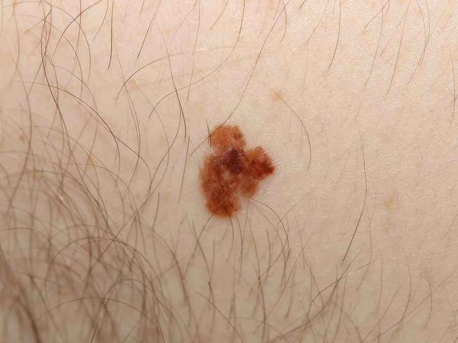
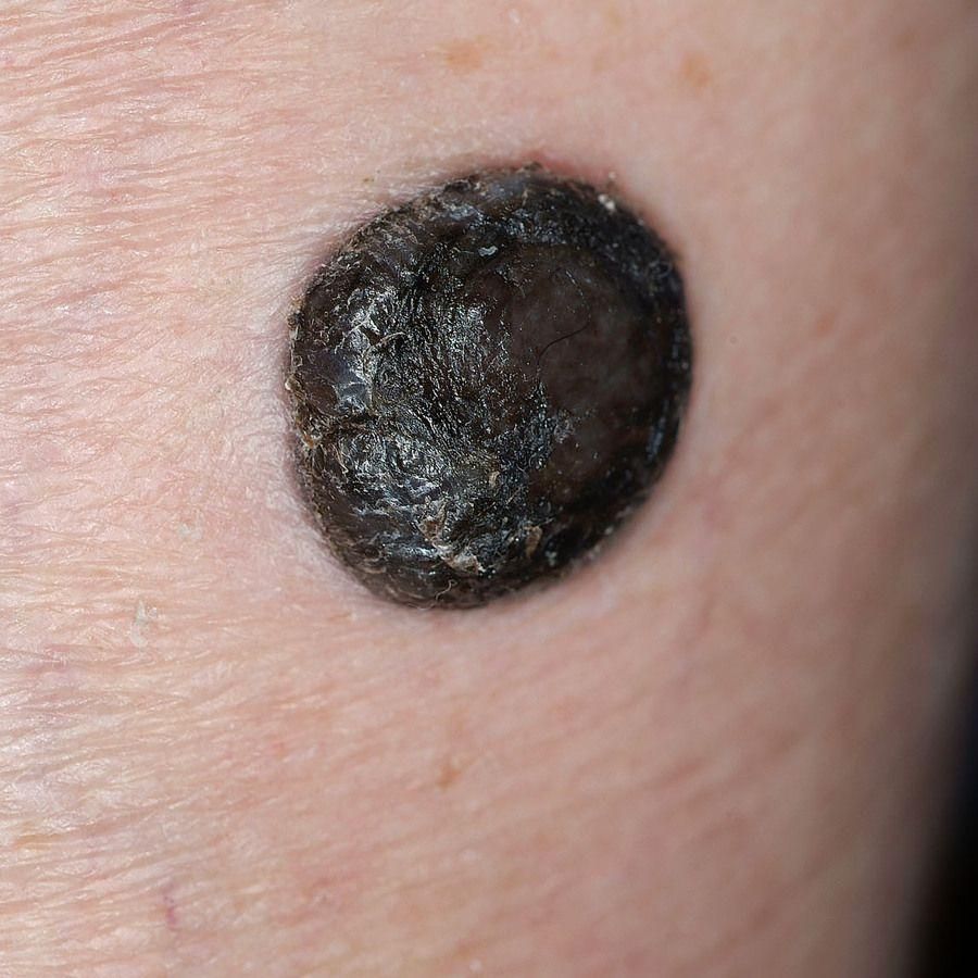
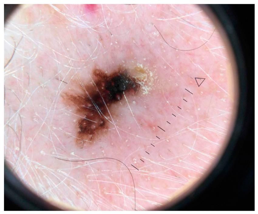
Early vs Advanced Melanoma
✓ Early melanoma
- Confined to the skin
- Usually treated surgically
- Excellent prognosis
✗ Advanced melanoma
- May spread to lymph nodes or internal organs
- Often requires specialist oncology treatment including immunotherapy or targeted therapy
- Early detection dramatically improves survival
Two clinical patterns: Fast-growing melanoma (often nodular) can be fatal within weeks — patients may not reach a doctor in time. Slow-growing melanoma (usually thin or superficial spreading) can take months or years before becoming dangerous, and is often indistinguishable from a normal mole. If you have regular skin checks, no one should die from it.
Types of Melanoma
1Superficial Spreading Melanoma (Most Common)
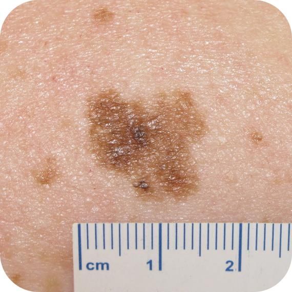
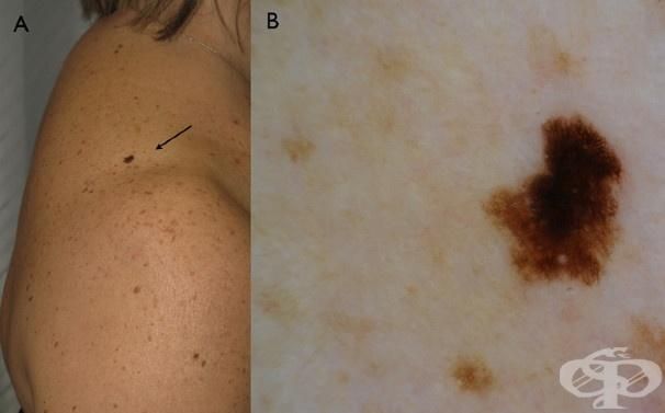
The most common type of melanoma in Australia. Characteristics: usually grows slowly in its early phase; begins as a flat irregular patch; colour is often uneven; borders become irregular; patients often notice a mole has changed over months or years.
2Nodular Melanoma
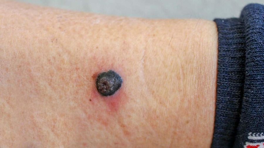
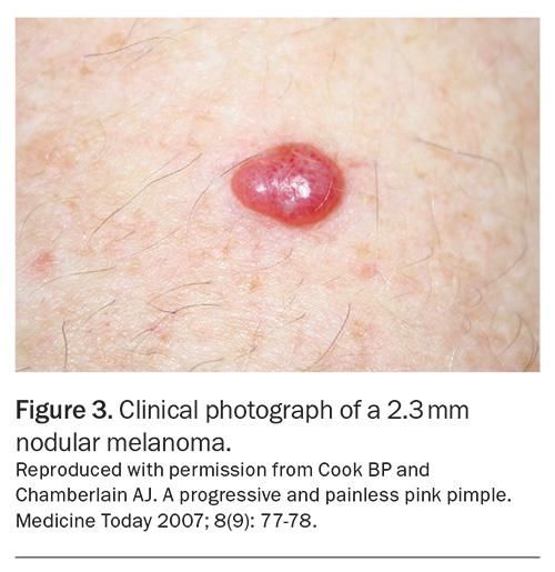
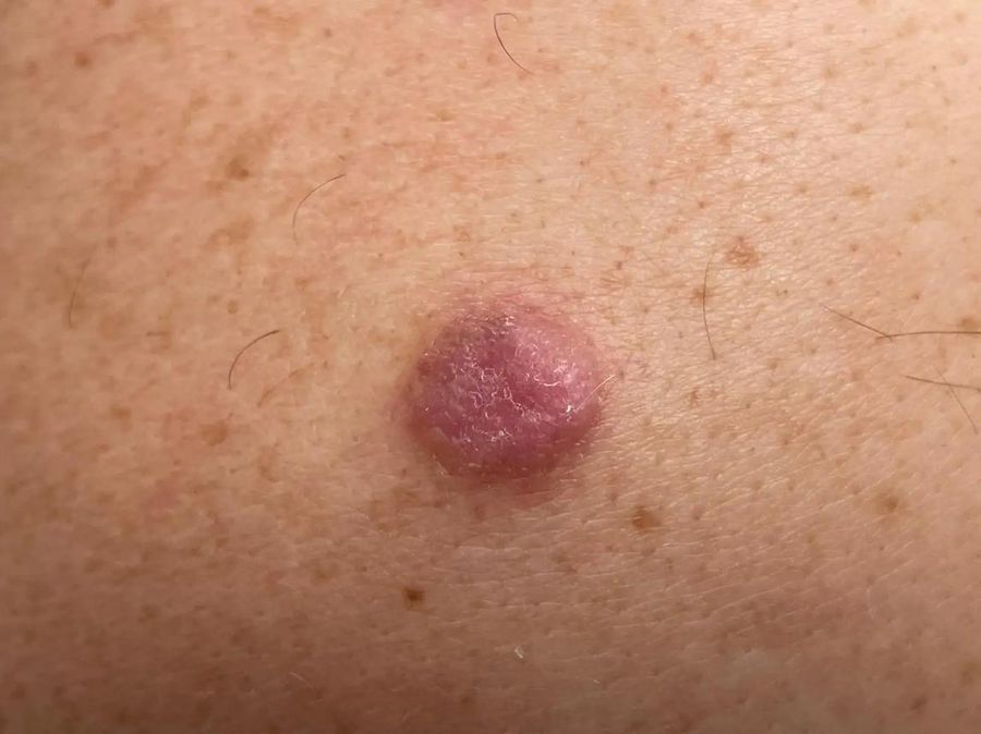
The most aggressive common melanoma. Characteristics: often grows vertically from the beginning; can develop over weeks to months; may be black, blue, brown, red or pink; frequently becomes thicker rapidly.
3Lentigo Maligna Melanoma
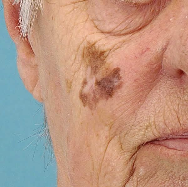
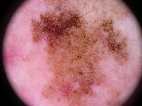
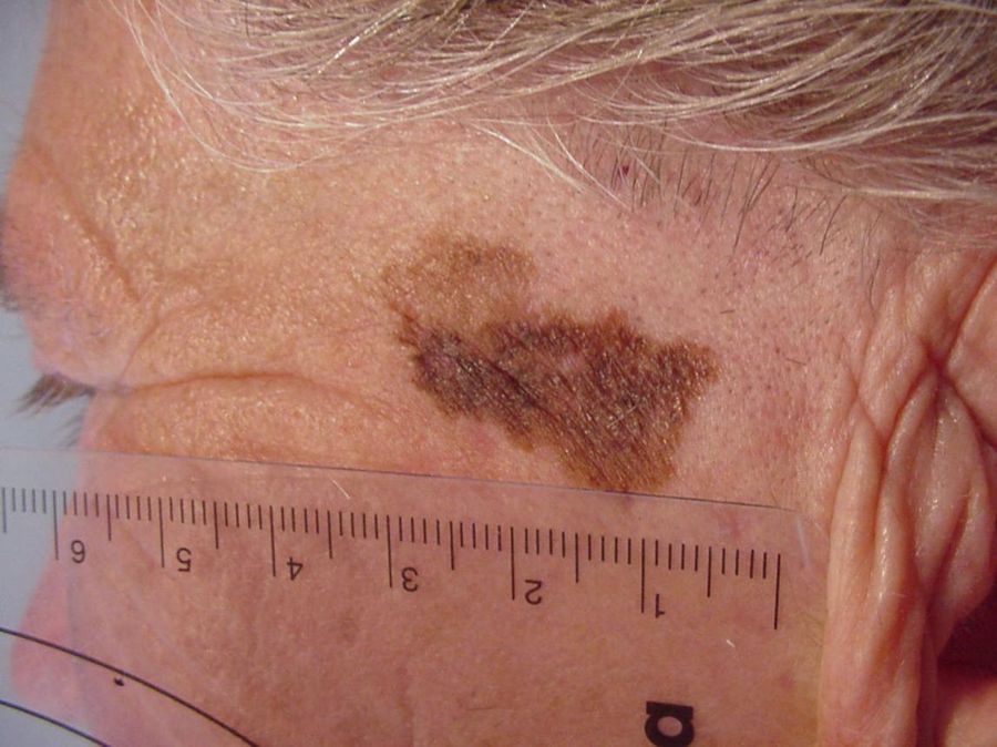
Very common on the face of older Australians with extensive sun damage. Usually develops from Lentigo Maligna, an early melanoma confined to the epidermis. Common locations: nose, cheeks, temples, forehead, ears. Often mistaken for an age spot.
4Acral Lentiginous Melanoma
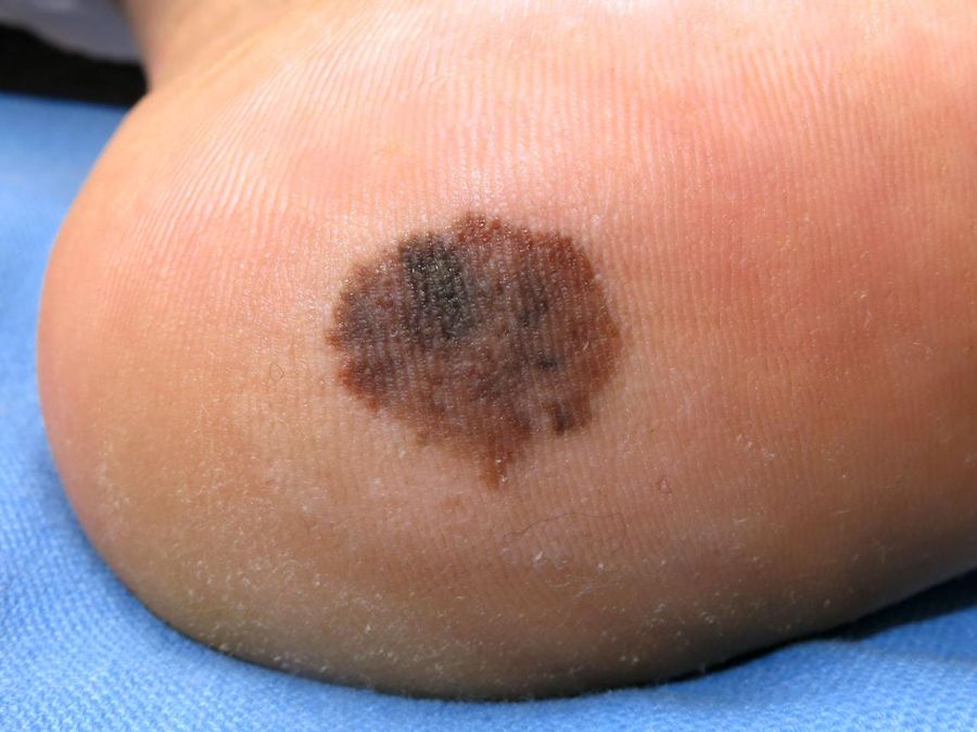
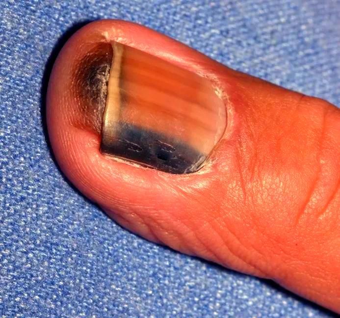
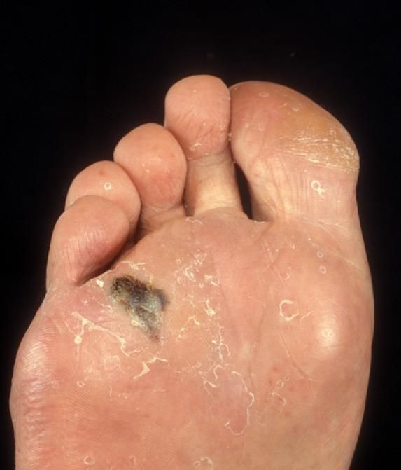
Occurs on the palms, soles, and under fingernails or toenails. Not related to sun exposure and can occur in all skin colours.
Basal Cell Carcinoma (BCC)
What is BCC?
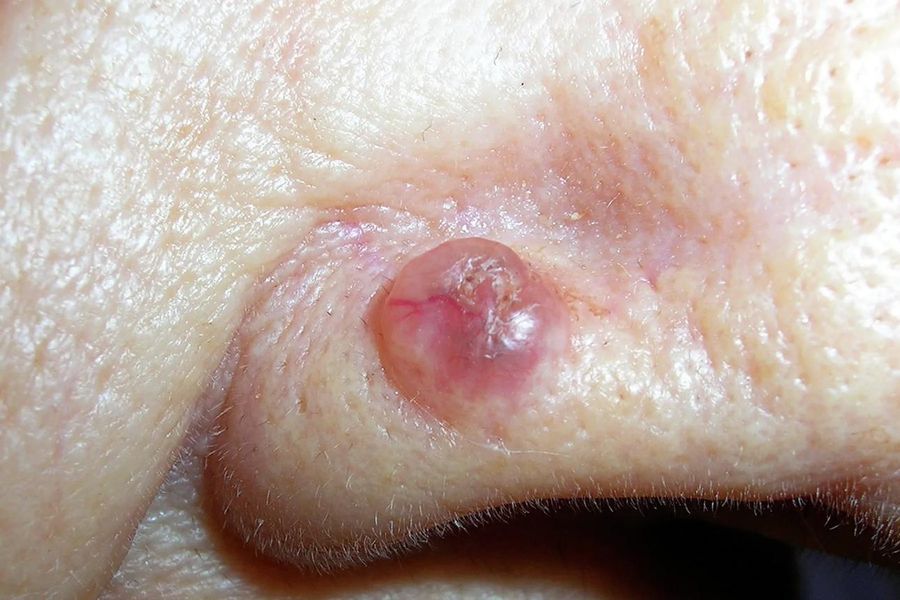
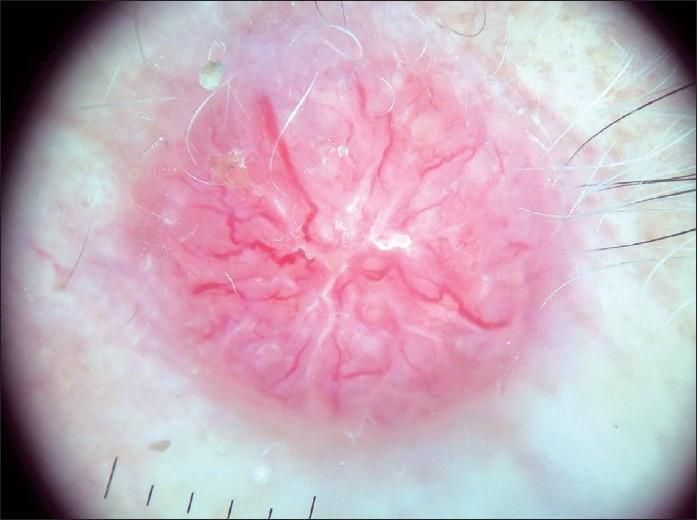
Basal Cell Carcinoma develops from basal cells in the lowest layer of the epidermis. It accounts for approximately 70% of non-melanoma skin cancers and is the most common cancer in Australia. Unlike melanoma, BCC grows slowly and rarely spreads to distant organs. However, untreated BCC can become locally destructive, invading cartilage, muscle, nerves and bone.
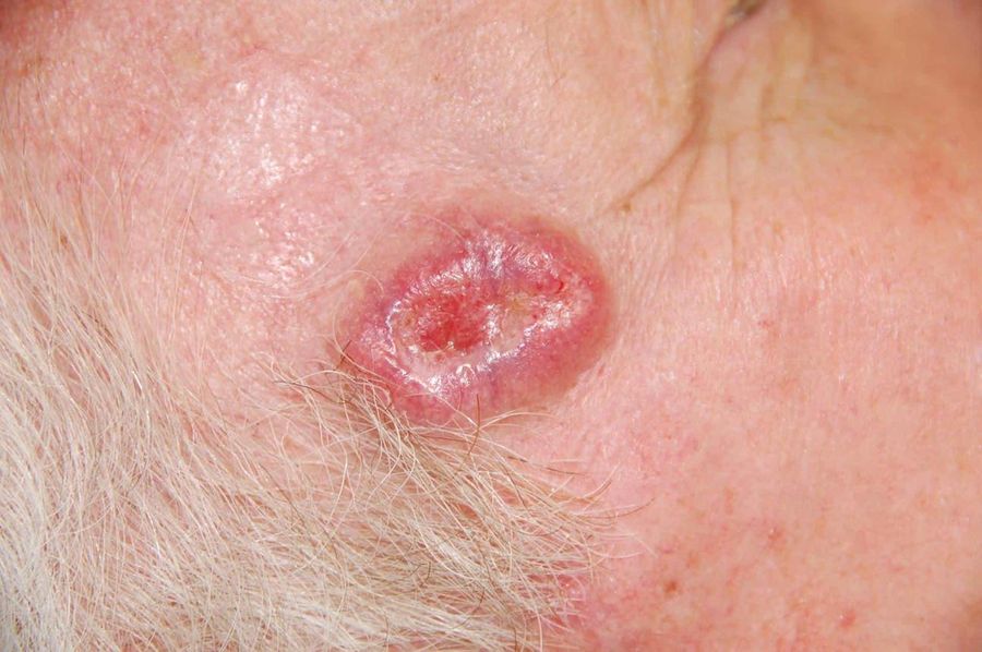
Why Does BCC Develop?
Usually due to:
- Long-term UV exposure
- Fair skin
- Increasing age
- Previous skin cancers
- Immunosuppression
Most BCCs occur on: nose, eyelids, ears, forehead, scalp, neck.
Different Types of BCC
1Nodular BCC — Most Common
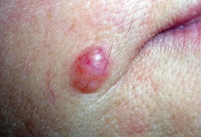
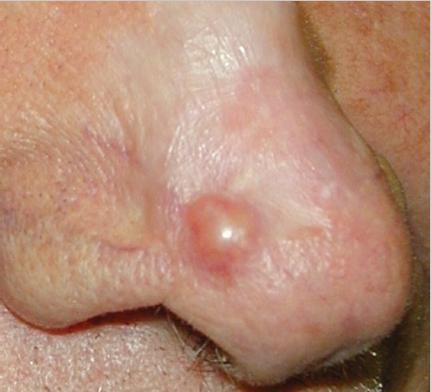
Features: pearly lump; visible blood vessels; bleeds easily; slowly enlarges.
2Superficial BCC
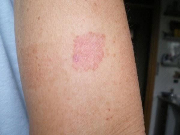
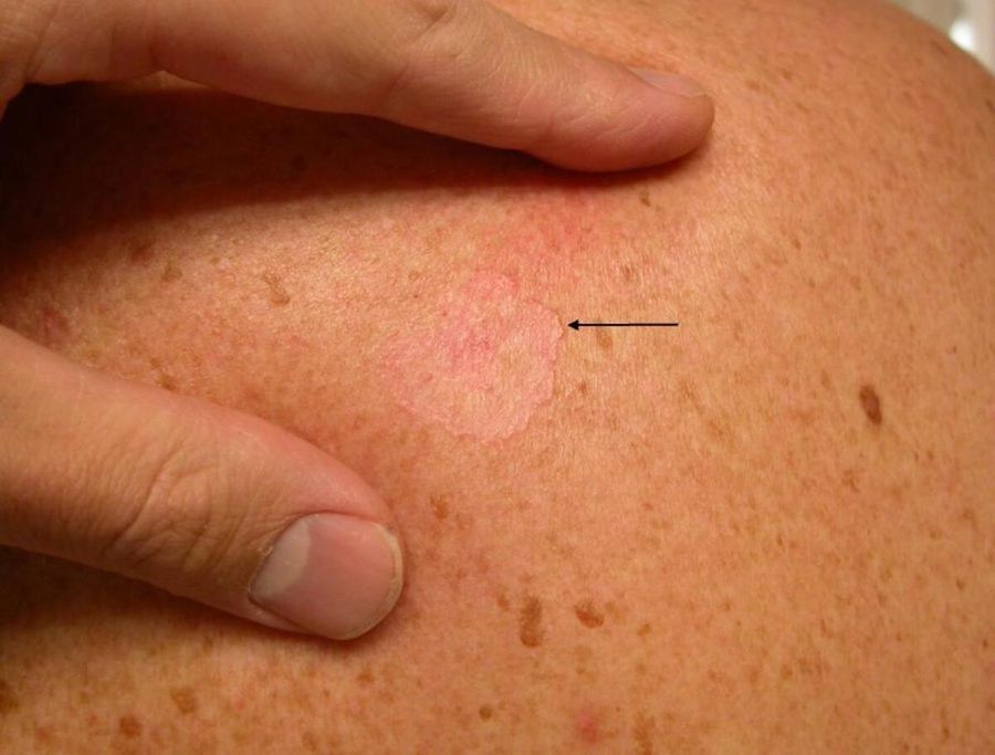
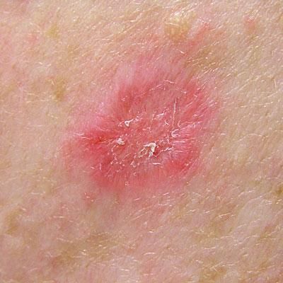
Common on trunk and shoulders. Looks like: red scaly patch; thin plaque; eczema-like lesion that never resolves.
3Pigmented BCC
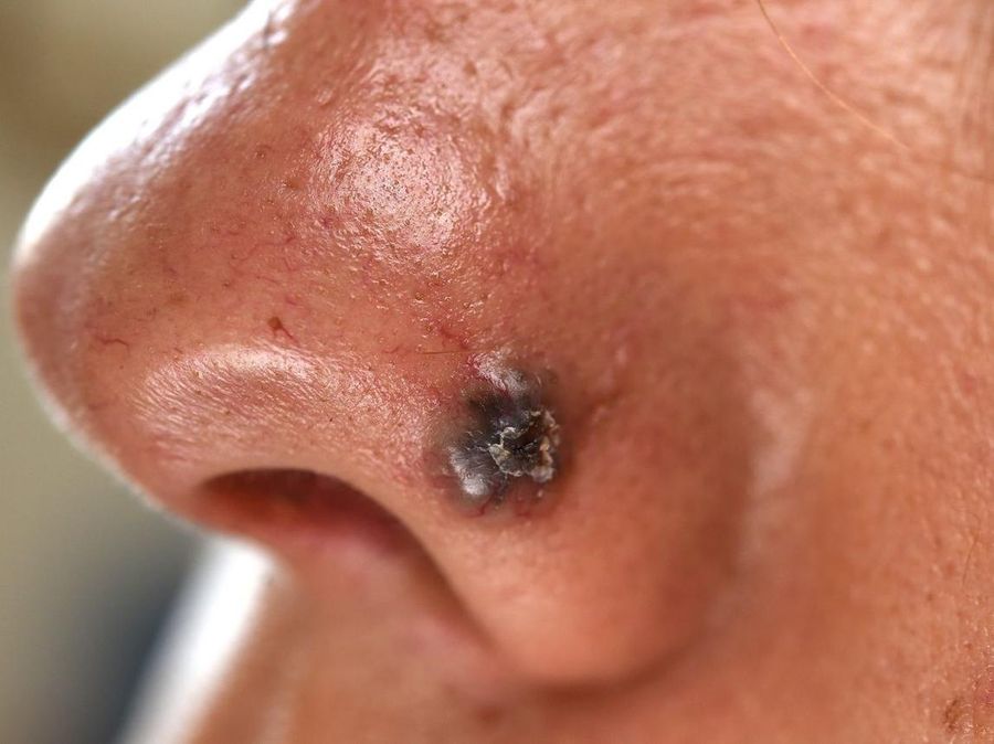
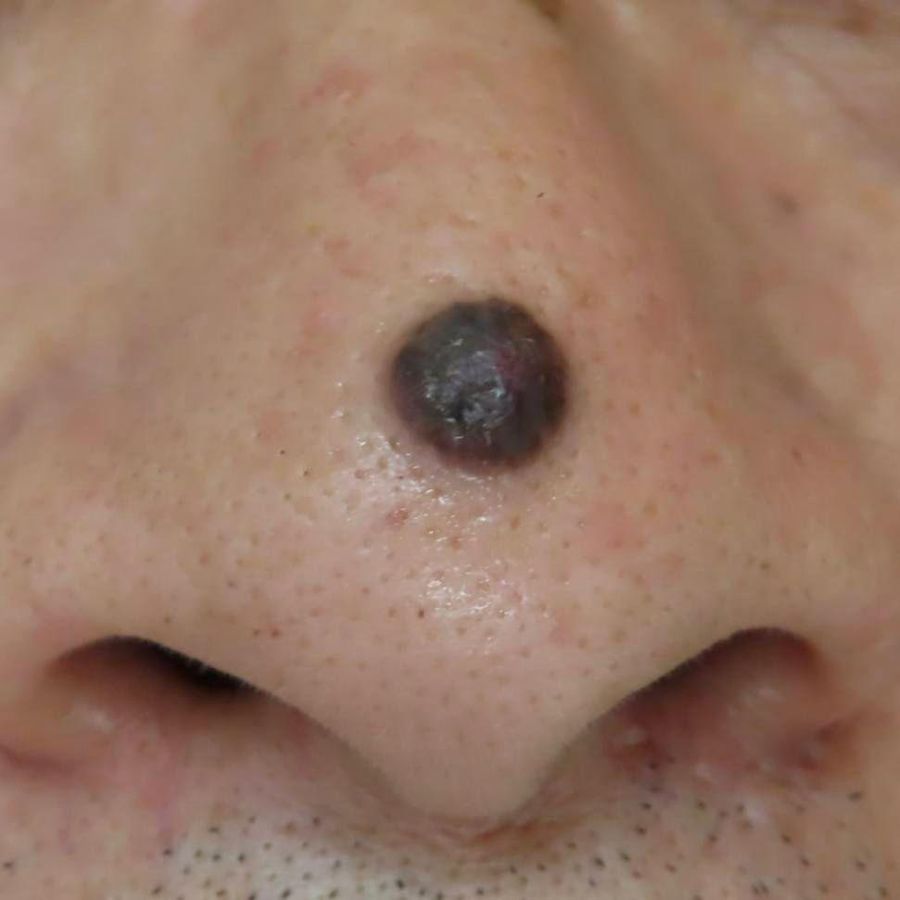
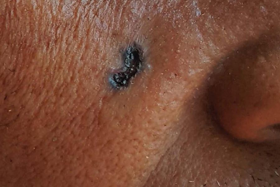
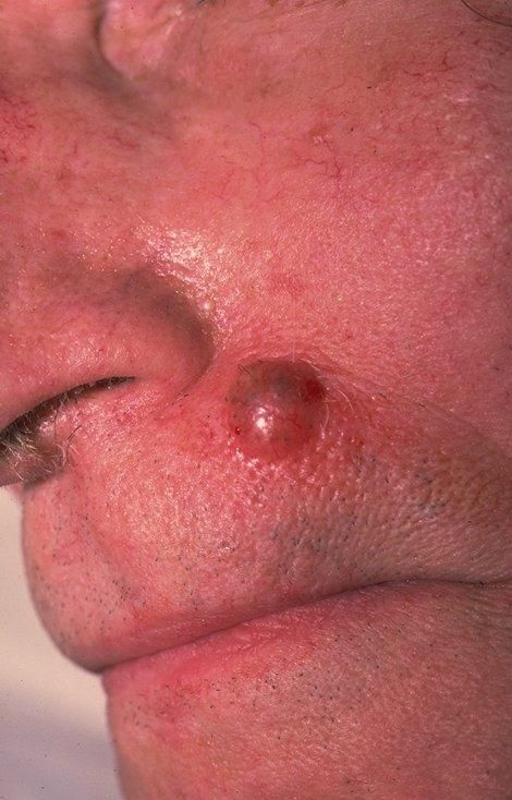
Contains brown, blue or black pigment. Often mistaken for melanoma. Requires dermoscopy to tell apart.
4Infiltrative / Morphoeic BCC
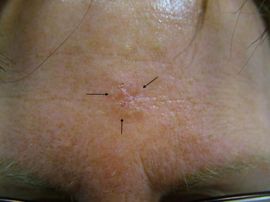
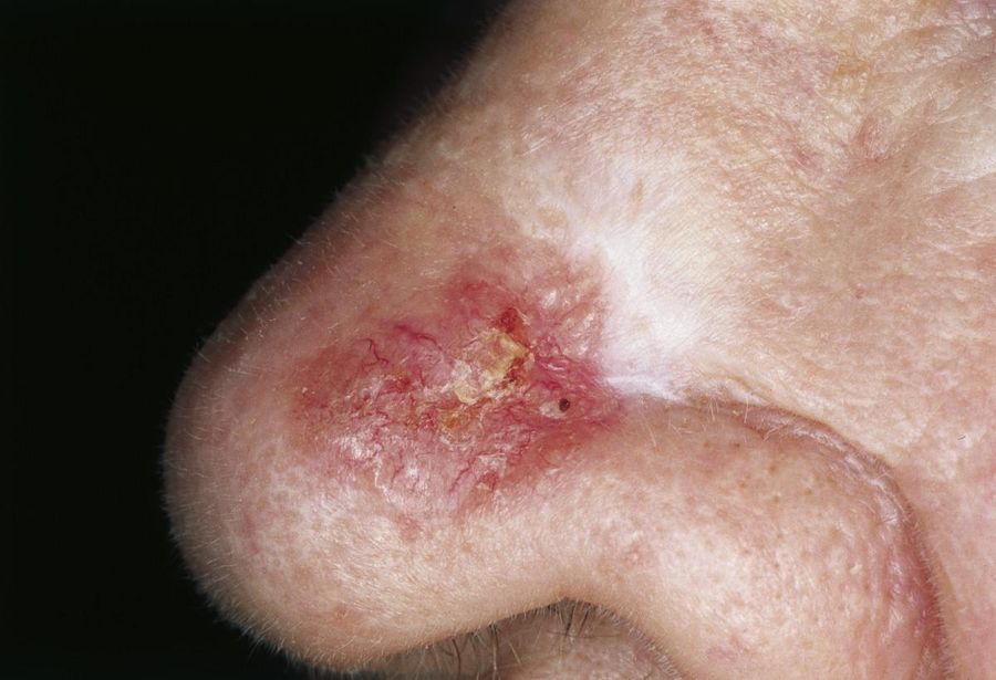
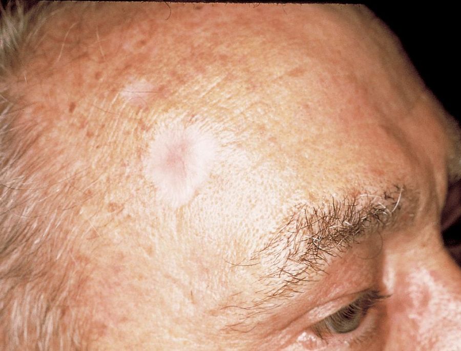
The most difficult subtype. Scar-like appearance; poorly defined borders; extends beneath normal skin; often requires more extensive surgery.
Warning Signs of BCC
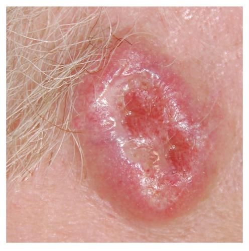
Patients should seek assessment if they notice:
- A shiny pearly bump
- A sore that repeatedly bleeds
- A crust that heals then returns
- A pink scaly patch persisting for months
- A scar-like white patch without prior injury
Is BCC dangerous?
BCC is usually not life-threatening. But untreated BCC may invade deeply, destroy cartilage, destroy eyelids, invade bone, and cause major cosmetic deformity. Early treatment is usually much simpler.
Squamous Cell Carcinoma (SCC)
What is SCC?
Squamous Cell Carcinoma develops from squamous cells in the upper epidermis. It accounts for approximately 30% of non-melanoma skin cancers in Australia. Unlike BCC, SCC has a greater ability to invade deeply and spread to lymph nodes and other organs, particularly when high-risk features are present.
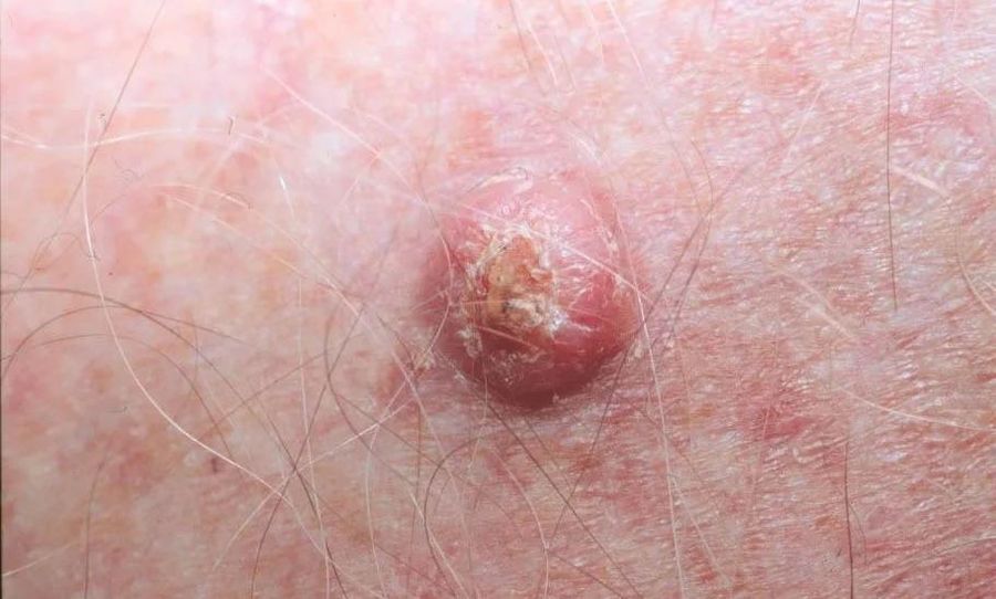
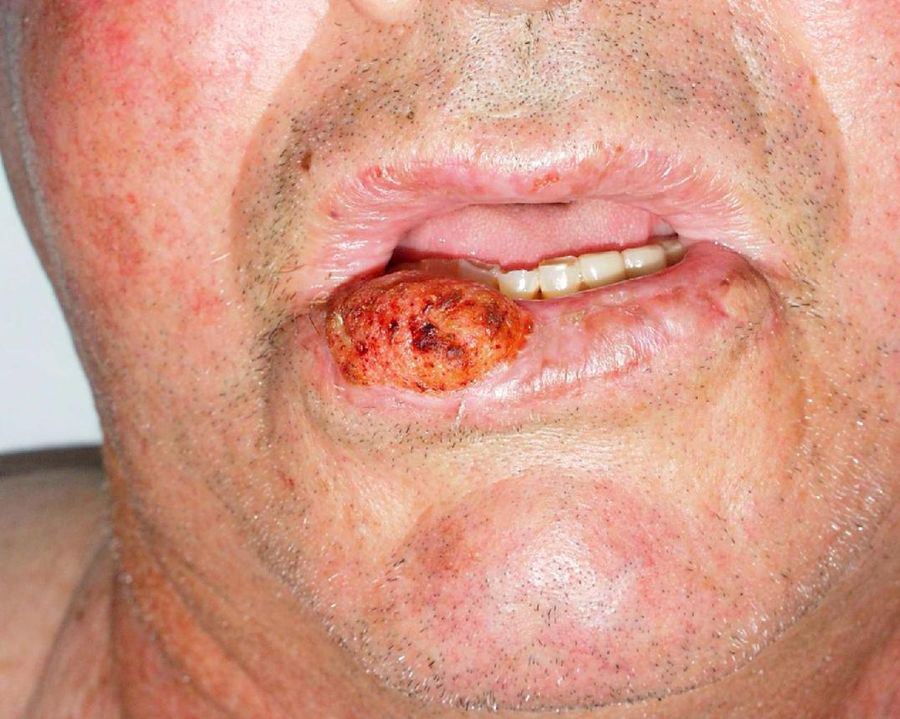
Why Does SCC Develop?
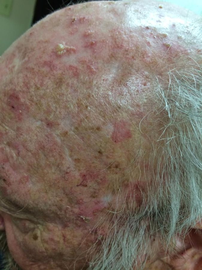
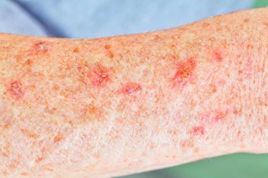
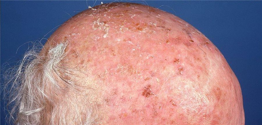
Usually develops after years of UV damage. Risk factors include:
- Chronic sun exposure
- Actinic keratoses (precancerous lesions)
- Fair skin
- Increasing age
- Immunosuppression
- Previous SCC
How SCC Appears
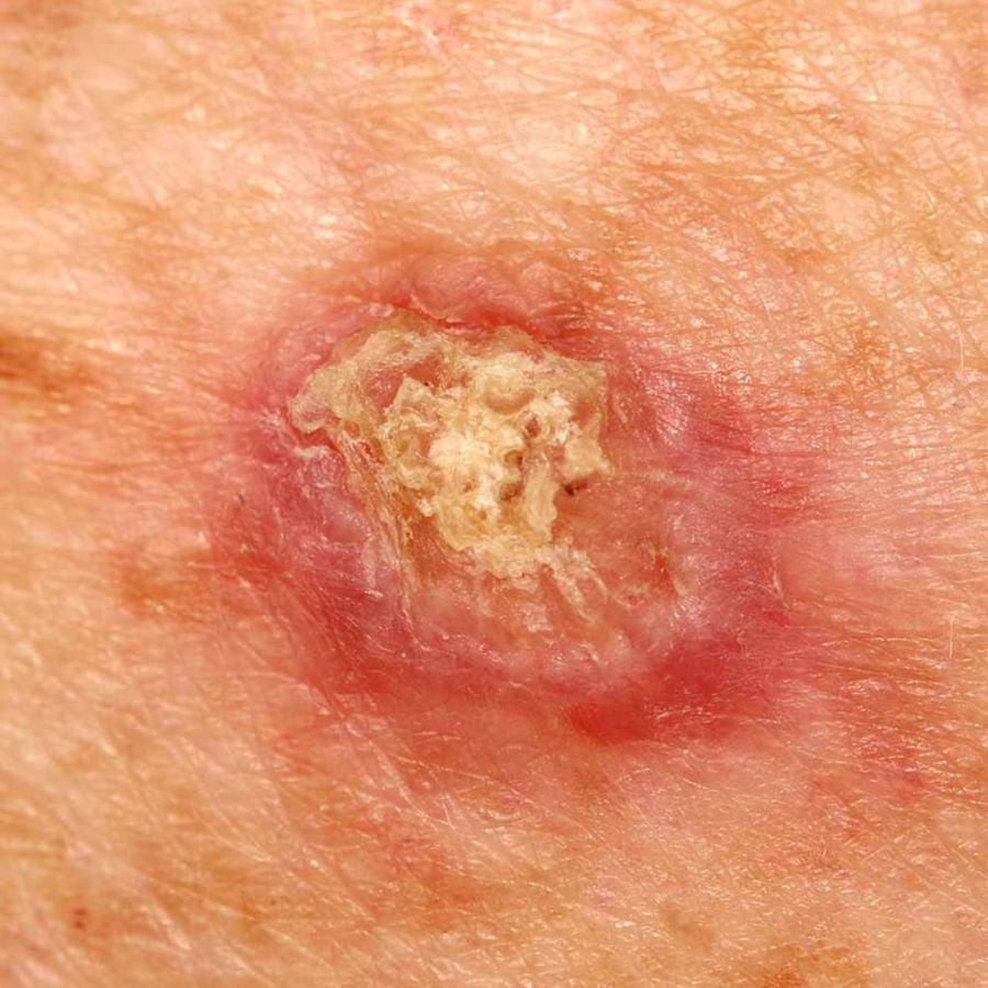
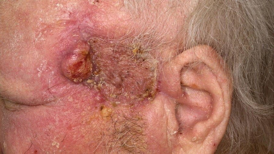
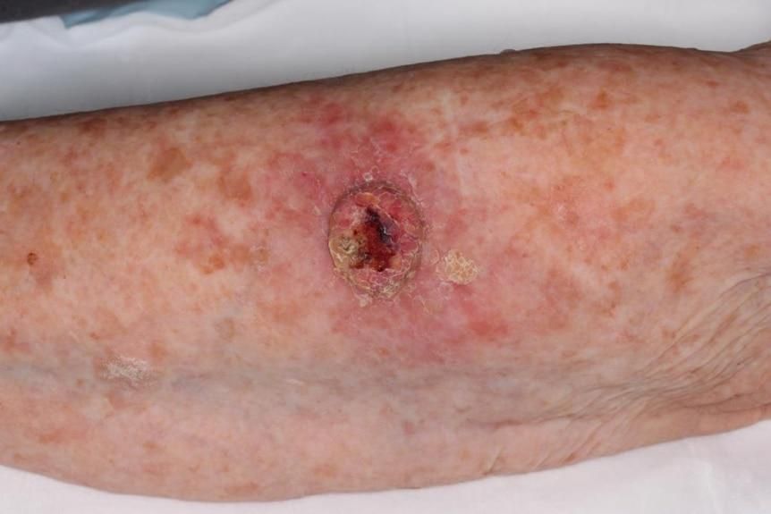
Common appearances include:
- Thickened scaly patch
- Tender red lump
- Wart-like growth
- Non-healing ulcer
- Rapidly enlarging crusted lesion
Common sites: scalp, face, ears, lips, hands, forearms, lower legs.
When SCC is Considered High Risk
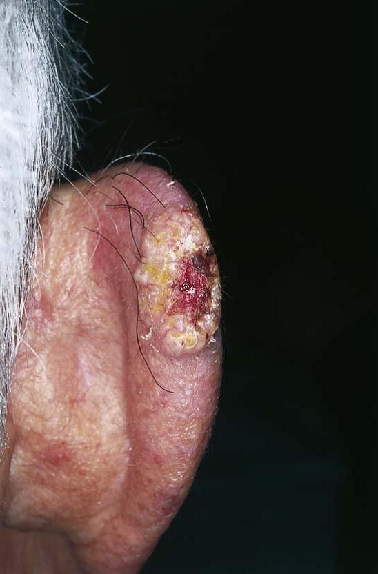
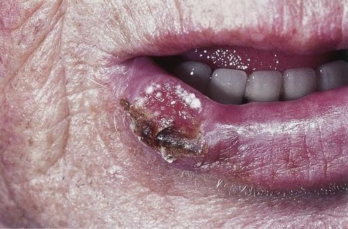
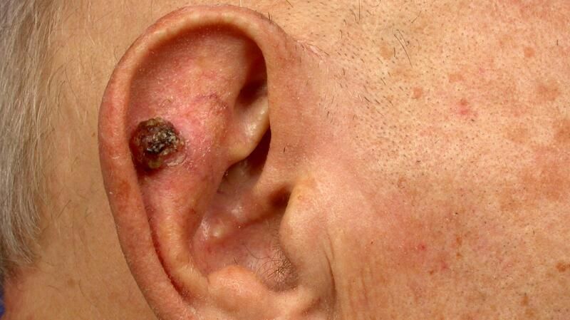
Higher-risk SCCs include lesions with:
- Greater thickness
- Poor differentiation
- Rapid growth
- Invasion around nerves (perineural)
- Lip or ear location
- Recurrence
- Immunosuppressed patients
These patients require closer follow-up.
Can SCC Spread?
Yes. Most SCCs are cured with surgery. However, untreated SCC may spread to lymph nodes, salivary glands, lungs and other organs. Risk depends on pathological features.
Actinic Keratosis — The Precursor of SCC
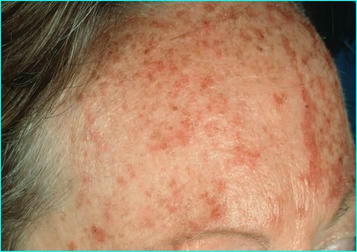
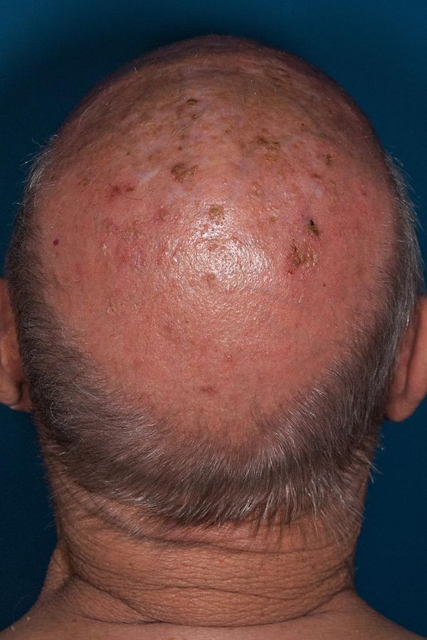
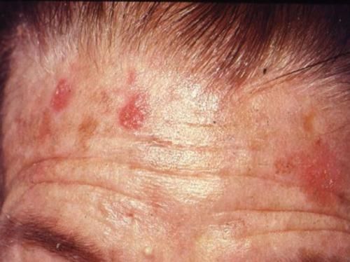
Actinic keratoses are precancerous lesions. They represent early UV-induced abnormal squamous cells. Some may remain stable, regress, or progress to SCC. This is why field treatment such as PDT is important for suitable patients.
Bowen Disease — Squamous Cell Carcinoma In Situ
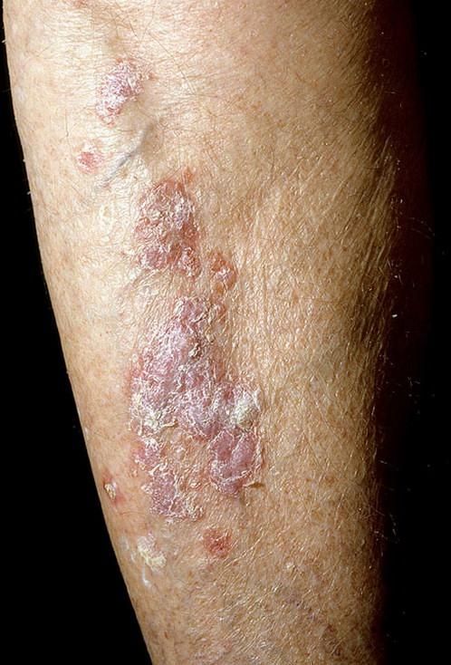
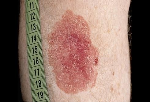
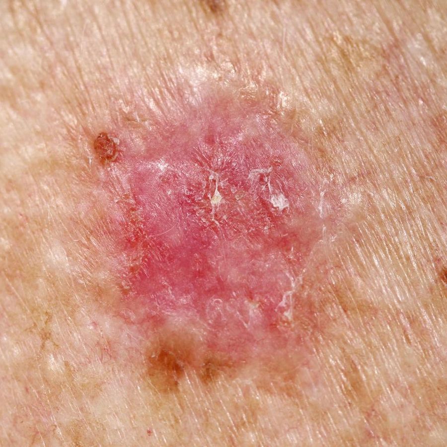
Bowen disease is the earliest stage of SCC. The cancer cells remain confined to the epidermis. Treatment options may include surgical excision, PDT, topical therapy, cryotherapy, or curettage. Early treatment prevents progression to invasive SCC.
Side-by-Side Comparison
| Feature | BCC | SCC | Melanoma |
|---|---|---|---|
| Cell of origin | Basal cells | Squamous cells | Melanocytes |
| Frequency | Most common | Second most common | Less common but most serious |
| Growth speed | Usually slow | Weeks to months | Variable |
| Can spread? | Rarely | Sometimes (lymph nodes / organs) | Yes (most likely to spread) |
| Typical appearance | Pearly lump | Scaly lump or ulcer | New or changing pigmented lesion |
| Main treatment | Surgery / PDT / topical | Surgery ± PDT (selected in situ) | Surgery ± oncology treatment |
When to Book a Check
Annual check: recommended for most Australian adults.
Every 6 months: if you have risk factors — fair skin, history of skin cancer, many moles, immunosuppression, or significant past sun exposure.
Immediately: if you notice a new or changing spot, a sore that won't heal, or a mole that looks different from the others.
Book a Skin Check Today
Early detection saves lives. Our doctors perform thorough dermoscopic examinations and arrange biopsy promptly if needed.
Book AppointmentDisclaimer: This page is for educational purposes only and does not constitute medical advice. Photographs are clinical reference images of disease states, not of identifiable individuals. If you have any concerns about a skin lesion, please consult a qualified medical practitioner.